AKI/CIN Prevention: CO₂ Angiography and the ROI of Renal Preservation
CO₂ angiography is a contrast-sparing strategy used in vascular procedures below the diaphragm to reduce, and in selected cases avoid, iodinated contrast media. This in-depth article is intended for vascular surgeons, interventional radiologists, and healthcare management teams who need to protect patients with chronic kidney disease, diabetes, or previous contrast reactions and assess the organizational and economic return of a renal-sparing platform.
CO₂ does not automatically eliminate every risk and does not replace individual clinical assessment. Its value lies in reducing iodine exposure through standardized protocols, with appropriate indications, contraindications, training, and outcome monitoring.
What are AKI and CIN, and why is prevention important?
AKI stands for “acute kidney injury,” meaning a sudden reduction in kidney function, assessed primarily through serum creatinine and urine output. AKI can prolong hospital stay, increase the risk of complications, and accelerate the deterioration of renal function, particularly in patients who already have CKD, or chronic kidney disease.
CIN, or contrast-induced nephropathy, is the term traditionally used to describe a worsening of renal function after exposure to iodinated contrast media. In modern clinical practice, a distinction is preferably made between a simple increase in creatinine temporally associated with contrast administration and kidney injury actually caused by contrast, often referred to as “contrast-associated AKI.” This distinction is important because infection, hypotension, embolization, and other conditions may also contribute to AKI.
Risk is not the same for every patient. Reduced eGFR, diabetes, advanced age, dehydration, heart failure, hypotension, and previous episodes of AKI increase vulnerability. International guidelines recommend identifying these factors before the procedure, using the lowest clinically appropriate volume of iodine, and adopting suitable renal protection measures.
How CO₂ angiography reduces exposure to iodinated contrast
CO₂ is a gaseous radiopaque contrast agent that, in appropriate vascular applications, allows visualization of the peripheral arterial circulation without introducing iodine into the body. It is particularly relevant in procedures below the diaphragm, such as selected peripheral revascularizations, arteriovenous fistulas for hemodialysis, and selected EVAR procedures.
The expected clinical benefit is a reduction in the total volume of iodinated contrast media. In a CO₂-first strategy, CO₂ is used as the primary imaging modality, while iodine is reserved for images or procedural steps in which it is truly necessary. In a CO₂-only strategy, applicable only to selected cases and vascular territories, the procedure may be performed without iodinated contrast.
Safety depends on patient selection and compliance with indications for use. CO₂ is not intended for arterial injection above the diaphragm and should not be regarded as a universal solution. A hospital protocol should define treatable anatomy, acquisition sequence, consumable management, image quality verification, and criteria for possible supplementation with small amounts of iodine.
What advantages does an automated renal-sparing platform offer?
Manual CO₂ injection may lead to variability in volume, pressure, timing, and image quality. An automated digital platform standardizes these parameters, makes the workflow more repeatable, and allows procedural data to be recorded. This helps the team compare cases, identify areas for improvement, and document the actual reduction in iodine use.
For a hospital, the benefit is not only technical. A renal-sparing system must integrate with the angiography suite, single-use consumables, clinical protocols, and training. Parameter traceability facilitates internal audits, quality assessments, and collection of indicators such as iodine volume per procedure, incidence of AKI at 48–72 hours, length of hospital stay, and readmissions.
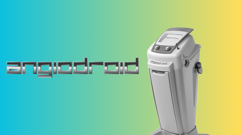
Angiodroid combines an automated CO₂ injector, a digital workflow, and Zero-Contrast training programs. The goal is not to promise complete elimination of risk, but to help centers reduce iodine exposure in a controlled, documented, and clinically consistent manner.
What is the expected clinical impact on reducing AKI?
The relationship between reducing iodine exposure and lowering the incidence of AKI is not identical in every population: it depends on baseline risk, the definition of AKI, the procedure, and other preventive measures. Available evidence on CO₂ angiography and contrast-sparing approaches suggests a potential benefit particularly in patients with CKD or other risk factors, but results should be interpreted according to study design and the clinical setting.
The KID Trial and real-world case series focused on renal preservation are examples of research designed to assess this issue in a structured manner. Before adopting a technology, a center should review endpoints, sample size, comparator, definition of AKI, iodine volume used, and duration of follow-up rather than relying solely on promotional percentages.
The most reliable way to estimate the local effect is to collect a baseline over at least several months and compare it with outcomes after implementation of the protocol. Indicators may include post-procedural AKI, worsening of eGFR, need for nephrology consultation, dialysis, iodine volume, length of hospital stay, reinterventions, and 30-day readmissions.
How to calculate the ROI of a CO₂ renal-sparing platform
ROI, or return on investment, should not be calculated solely on the price of the injector. The assessment should compare the total cost of adoption with the avoided costs and the clinical and organizational benefits observed at the center.
- Define the baseline: number of eligible procedures, average iodine volume, AKI rate, hospital stay, readmissions, and consumable costs.
- Estimate the benefit: expected reduction in iodine use, AKI episodes, length of hospital stay, and procedures postponed or modified because of renal risk.
- Account for the investment: system, consumables, training, maintenance, staff time, and workflow integration.
- Measure the outcome: after 6–12 months, compare indicators with the baseline and calculate the payback period, meaning the time required to recover the investment.
A simple formula is: ROI = (annual economic benefits − total annual cost of the solution) / total annual cost of the solution × 100. Economic benefits should be documented locally: the cost of an AKI episode varies according to severity, length of hospital stay, monitoring, consultations, dialysis, and readmissions. For this reason, it is not appropriate to assign a universal ROI without data from the individual hospital.
As an example of a business case, a center may estimate the annual value of each avoided AKI episode and multiply it by the observed reduction, adding the savings generated by lower iodine volumes and fewer hospital days. The result is a scenario model, not a guarantee of savings, and should be validated using actual administrative and clinical data.
Which patients and procedures may benefit the most?
A CO₂-first approach is most relevant when renal risk is high and the procedure involves an appropriate vascular territory. Cases to consider include patients with reduced eGFR, diabetes, progressive CKD, previous events associated with iodinated contrast, or documented allergy, as well as peripheral procedures and selected complex aortic interventions.
The decision should be multidisciplinary. The interventional radiologist, vascular surgeon, anesthesiologist, and, when necessary, nephrologist should balance image quality, urgency, anatomy, embolic risk, cardiopulmonary function, and the possibility of completing the procedure with CO₂, low-volume iodine, or other imaging modalities.
How to implement a measurable Zero Contrast pathway
An effective pathway starts with case selection, not with purchasing the device. The center should establish a working group, approve a protocol, train staff, and introduce a checklist with inclusion, exclusion, and conversion criteria for iodinated contrast when clinically necessary.
Practical training through workshops, live cases, and mentorship reduces the learning curve and helps maintain adequate image quality. Monitoring should be continuous: each procedure should record imaging modality, iodine volume, radiation dose, duration, complications, and renal outcomes.
To learn more about evidence, adoption protocols, and evaluation opportunities at your center, request a clinical consultation and a demo of the CO₂ renal-sparing platform. The material can help the team estimate indications, KPIs, and economic sustainability based on its own procedural volume.
Frequently Asked Questions
Does CO₂ angiography eliminate the risk of AKI?
No. CO₂ angiography can reduce exposure to iodinated contrast and therefore contribute to the prevention of contrast-associated kidney injury, but it does not eliminate all causes of AKI. Risk also depends on hypotension, embolization, infection, dehydration, and pre-existing conditions.
Is CO₂ an alternative to iodinated contrast in every angiographic procedure?
No. CO₂ is mainly used for vascular procedures below the diaphragm and has specific indications and contraindications. The choice between CO₂, low-volume iodine, or other techniques depends on anatomy, the procedure, and the patient’s clinical condition.
What data are needed to estimate hospital ROI?
At a minimum, the center needs the number of eligible procedures, average iodine volume, AKI incidence, hospital stay, readmissions, and the costs of technology, consumables, and training. ROI should be calculated using local data and verified after implementation, ideally by comparing the baseline period with the period of use.
Why choose an automatic injector instead of manual CO₂ injection?
An automatic injector can standardize volume, pressure, timing, and parameter recording, reducing operator-dependent variability. Training and an appropriate protocol nevertheless remain essential for using CO₂ safely and correctly.
Which clinical indicators should a center monitor?
Key indicators include AKI at 48–72 hours, changes in eGFR, iodine volume per procedure, complications, length of hospital stay, readmissions, and need for dialysis. It is also useful to include radiation dose, procedure duration, and the conversion rate from CO₂ to iodine.
Clinical note: this content is for informational purposes only and does not replace guidelines, specialist assessment, or the medical device instructions for use.